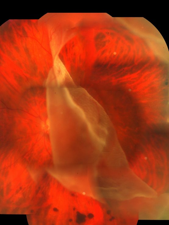
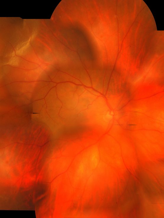
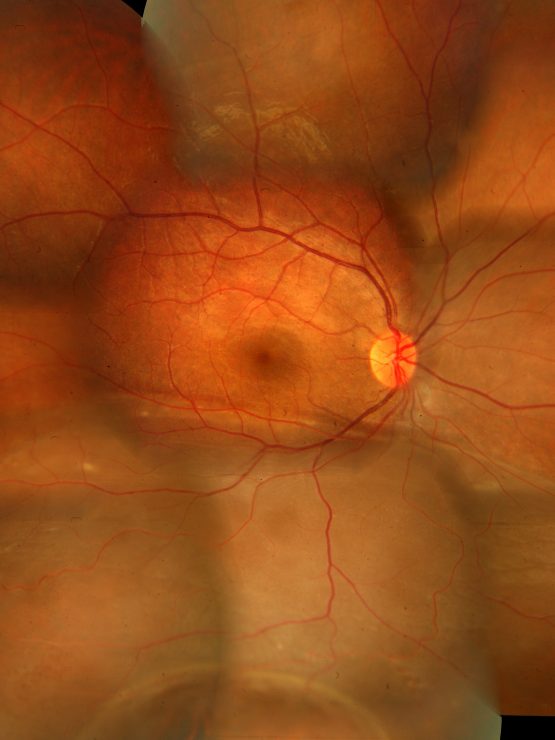
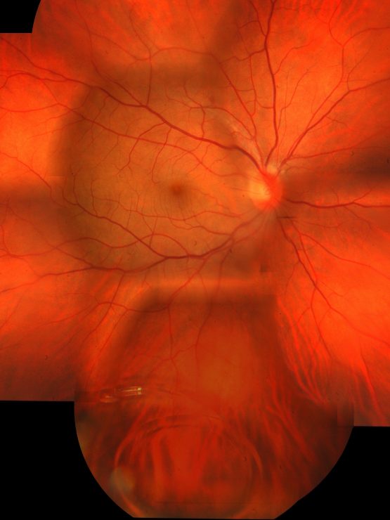
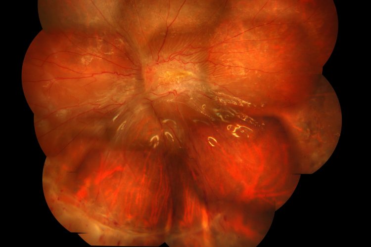
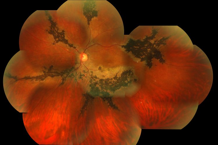
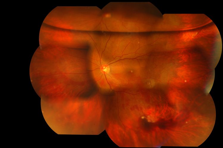
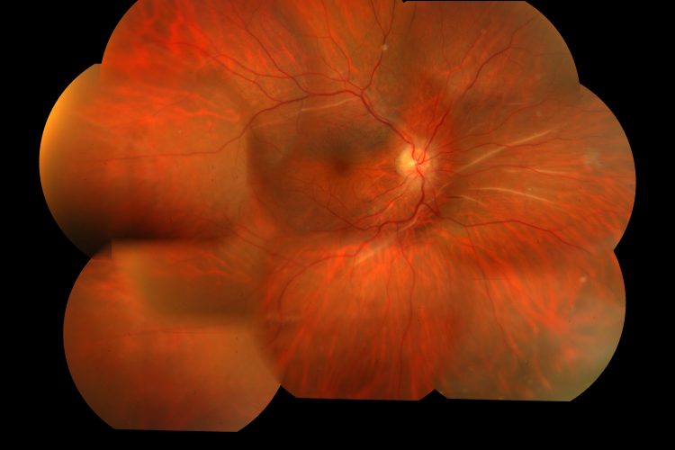
Advanced Retinal Photography and Eye Retina Images for Optimal Eye Health
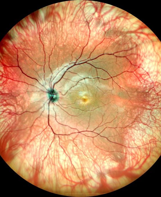
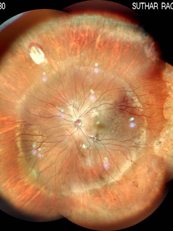
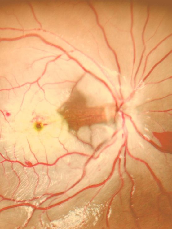
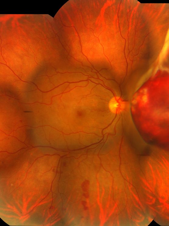
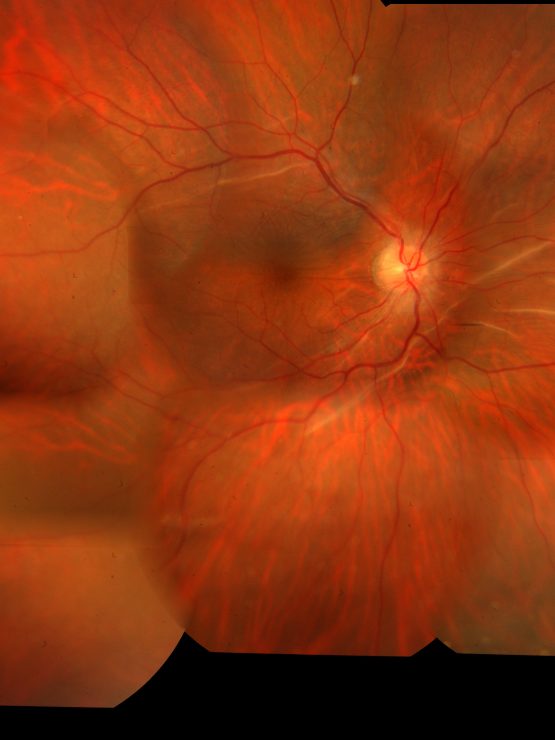
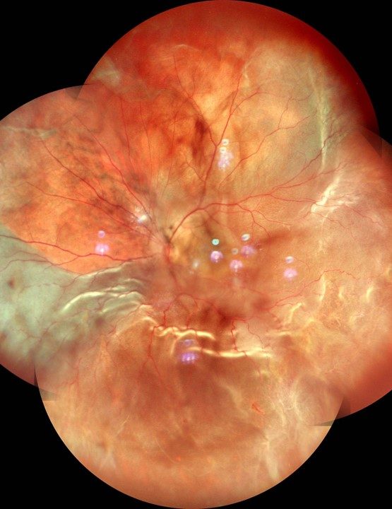
Retina Foundation offers state-of-the-art retinal photography and detailed eye retina images to support optimal eye health. By utilising cutting-edge technology, we provide comprehensive monitoring of your eye anatomy and ocular health.
Importance of Retinal Photography and Eye Health
The Significance of Retinal Imaging for Eye Health
Retinal imaging is essential for maintaining good eye health. Techniques like fundus photography, OCT imaging, fluorescein angiography, and digital retinal photography provide detailed views of the retina. These methods help in early diagnosis and management of various eye conditions. At Retina Foundation, advanced technology such as 3D eye imaging offers high-resolution images that aid in accurate monitoring and treatment planning.
By capturing clear images of the eye retina, these technologies allow ophthalmologists to detect problems early. This early detection is vital because many retinal diseases progress without noticeable symptoms until significant damage has occurred. Regular retinal imaging can prevent severe vision loss by identifying issues before they become critical.
Common Conditions Detected by Retinal Imaging
Retinal imaging helps diagnose several common eye conditions:
- Central Retinal Artery Occlusion (CRAO): CRAO involves the blockage of the central retinal artery, leading to sudden vision loss. Early detection through retinal imaging can help manage this condition effectively.
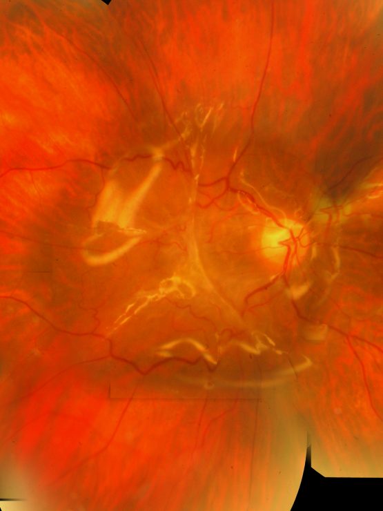
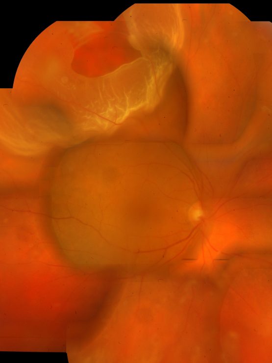
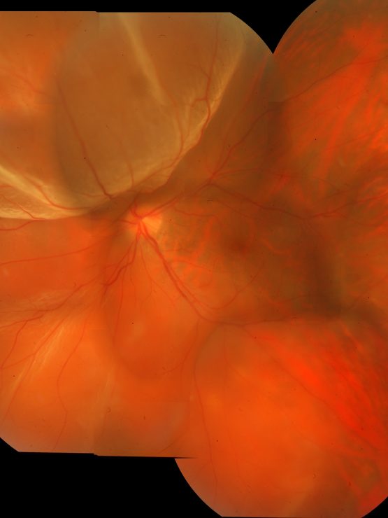
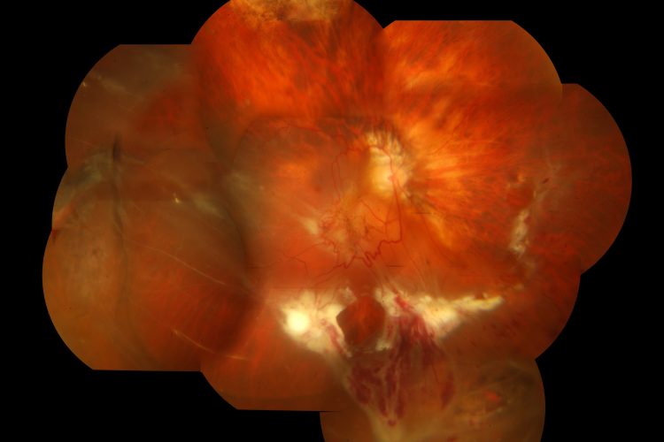
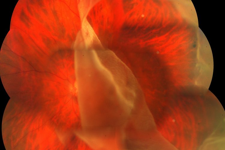
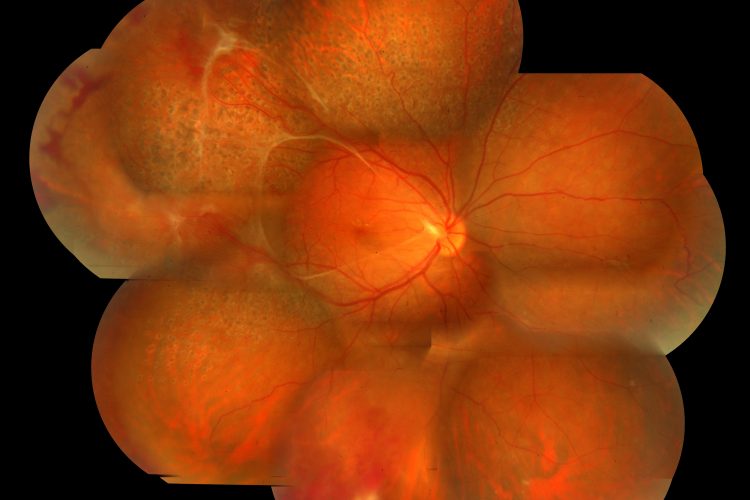
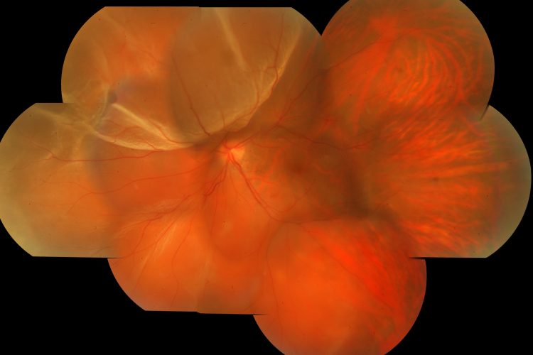
- Retinal Detachment: This occurs when the retina separates from its underlying tissue. Prompt treatment is crucial to prevent permanent vision loss. Retinal images help identify tears or holes that may lead to detachment.
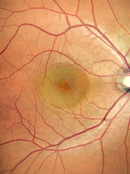
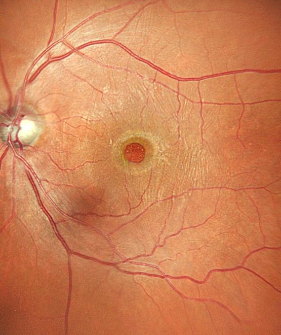
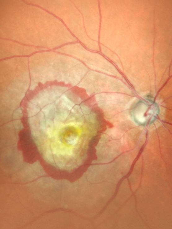
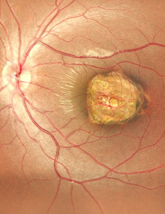
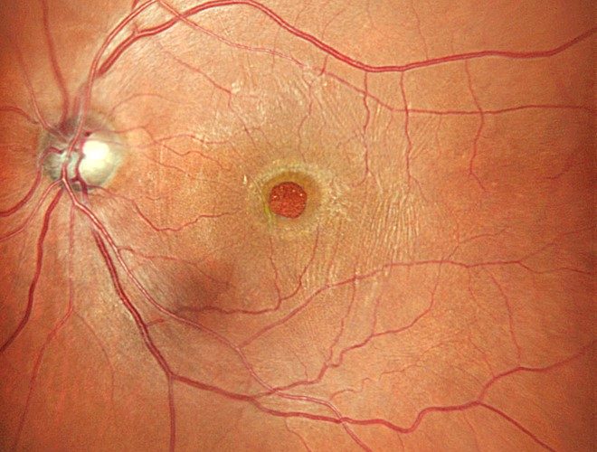
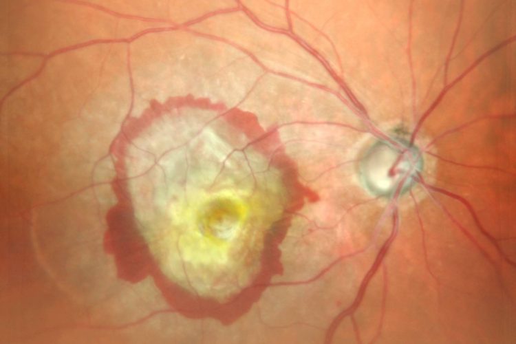
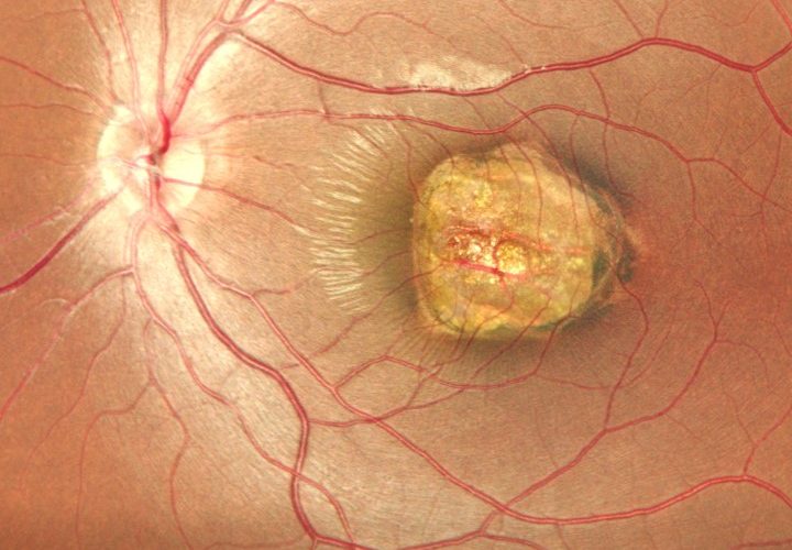
- Macular Hole: A small break in the macula can cause blurred and distorted vision. Retinal imaging helps detect macular holes early on, allowing for timely surgical intervention.
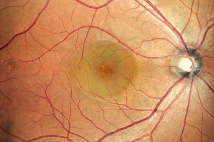
- Central Serous Retinopathy (CSR): CSR involves fluid accumulation under the retina, causing visual disturbances. Imaging techniques like OCT provide detailed pictures that aid in diagnosing and monitoring CSR.
- Choroidal Melanoma: This type of cancer affects the choroid layer beneath the retina. Early detection through imaging is essential for effective treatment.
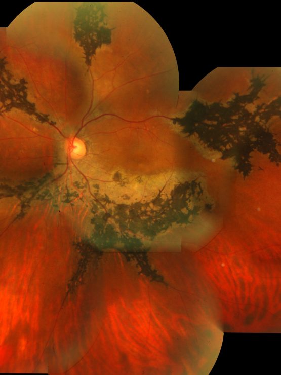
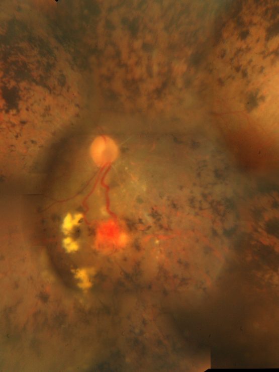
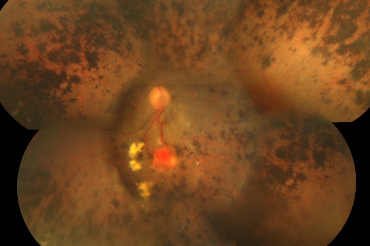
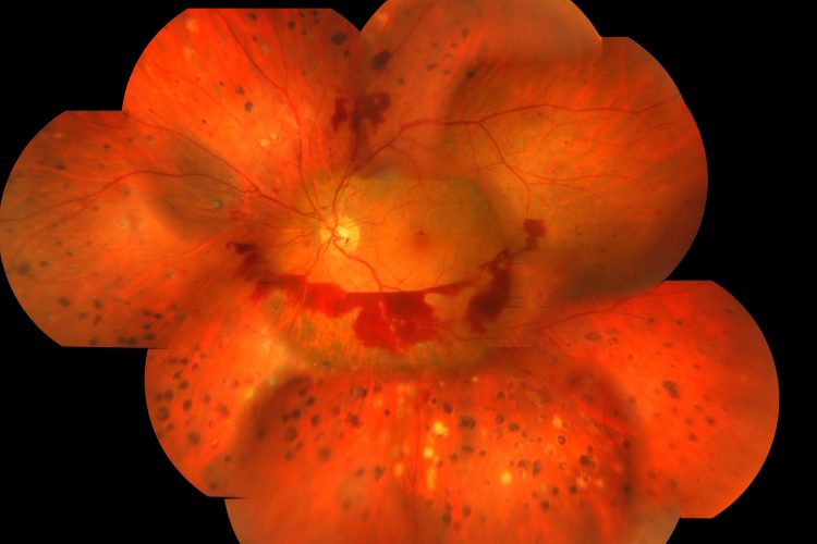
- Retinitis Pigmentosa: A group of genetic disorders affecting how the retina responds to light. Detailed retinitis pigmentosa images assist in understanding disease progression.
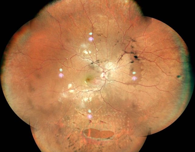
- Diabetic Retinopathy: High blood sugar levels can damage retinal blood vessels, leading to diabetic retinopathy. Regular imaging helps monitor changes and guide treatment decisions.
Benefits of Regular Retina Check-ups
Routine retina check-ups offer numerous benefits:
- Preventive Care: Regular check-ups enable early detection of potential issues before they escalate into severe problems.
- Customized Patient Care at Retina Foundation: Personalized care plans are designed based on individual needs and conditions detected during check-ups.
- Maintaining Optimal Eye Health: Consistent monitoring ensures any changes in eye health are promptly addressed.
Regular visits to specialists at Retina Foundation ensure comprehensive evaluations using state-of-the-art equipment, ultimately preserving your vision health.
Advanced Technology in Retinal Imaging
The advancements in retinal photography have revolutionized eye care:
Traditional Methods vs Modern Technological Advancements: Traditional methods relied heavily on basic ophthalmoscopes with limited diagnostic capabilities. In contrast, modern techniques such as OCT (Optical Coherence Tomography) provide cross-sectional images offering unparalleled detail.
Cutting-edge Equipment at Retina Foundation: Utilizing advanced tools like 3D imaging systems enhances diagnostic accuracy and patient outcomes compared to older methods.
These technological innovations not only improve diagnostic precision but also enhance patient experience by providing quicker results with minimal discomfort.
Real-life Stories: How Retinal Imaging Saved Sight
Real-life experiences highlight the importance of regular retinal imaging:
- A patient experiencing sudden vision loss was diagnosed with central retinal artery occlusion during a routine check-up at Retina Foundation. Early intervention helped restore partial vision.
- Another case involved detecting a macular hole early through OCT scans, allowing for successful surgical repair before significant vision impairment occurred.
- Diabetic patients benefiting from regular screenings had their diabetic retinopathy managed effectively through timely treatments guided by detailed imagery.
These testimonials underscore how proactive monitoring using advanced retinal photography techniques can save sight and significantly improve quality of life.
Incorporating regular retinal photography into your healthcare routine is essential for maintaining good eye health and preventing serious conditions from progressing undetected.
Retinal Detachment and Its Variants
Types of Retinal Detachment
Inferior RD
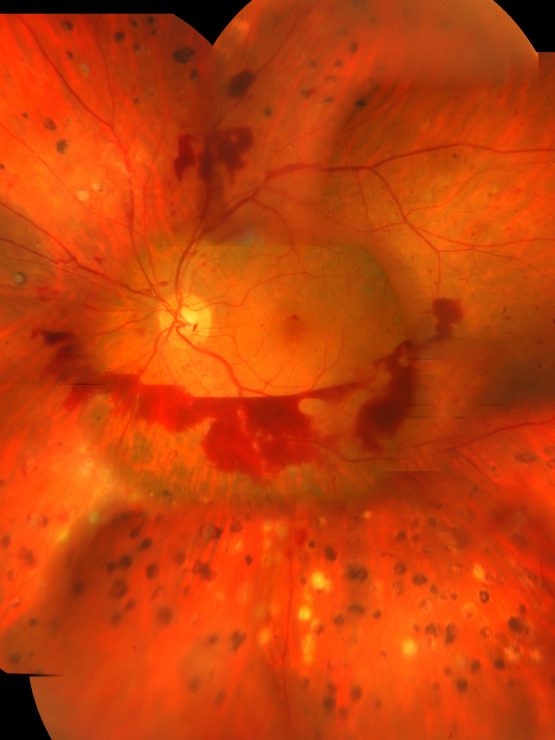
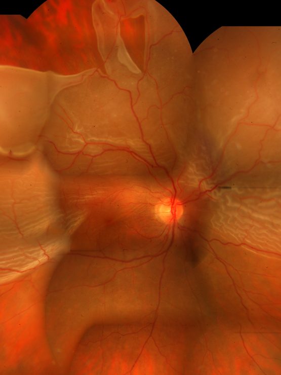
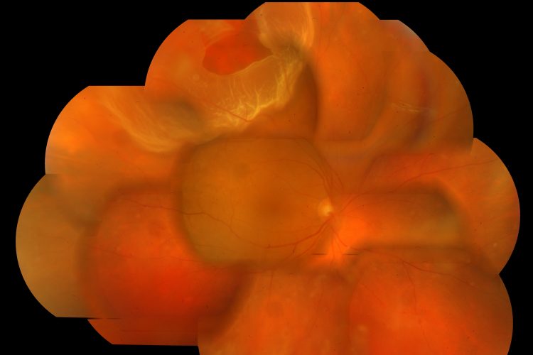
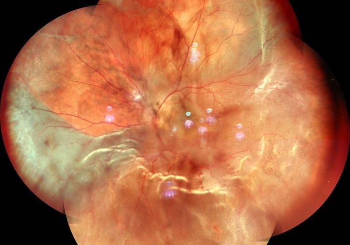
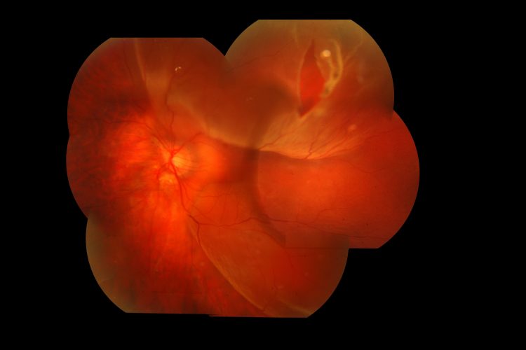
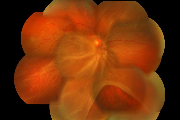
Inferior retinal detachment happens when the retina pulls away from its base in the lower part of the eye. Causes include aging, trauma, or severe myopia. Symptoms like seeing flashes of light, floaters, or a shadow over vision are common. Advanced imaging techniques such as Optical Coherence Tomography (OCT) can spot inferior RD early, allowing for quick treatment.
Superior RD with Large Tear
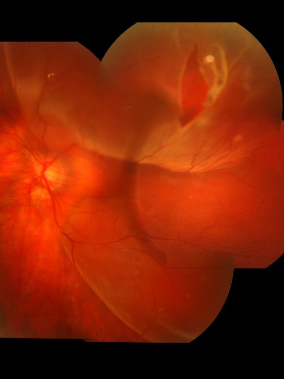
A superior retinal detachment with a large tear is serious and needs immediate attention to avoid permanent vision loss. This condition often presents sudden vision changes or an increase in floaters. Prompt treatment through surgery or laser therapy is essential to restore vision.
Giant Retinal Tears
Giant retinal tears cover more than 90 degrees of the retina’s circumference and are more complex than other detachments. High-resolution imaging techniques are crucial for identifying these tears accurately and planning the right surgical interventions.
Identifying Symptoms Through Detailed Imagery
Symptoms Detection
Common symptoms of retinal detachment include:
- Flashes of light
- Floaters
- Shadowy vision
These symptoms can be detected through detailed ocular imagery, which provides a clear view of the retina’s condition.
Imaging Techniques
Various imaging techniques help identify retinal detachment symptoms accurately:
- Optical Coherence Tomography (OCT): Provides high-resolution cross-sectional images.
- Fundus Photography: Captures detailed images of the retina.
- Fluorescein Angiography: Highlights blood flow issues in the retina.
Treatment Options Available at Retina Foundation
Vitrectomy
Vitrectomy is a surgical procedure used for severe cases of retinal detachment by removing vitreous gel and replacing it with a saline solution or gas bubble. This helps reattach the retina and restore vision.
Laser Therapy
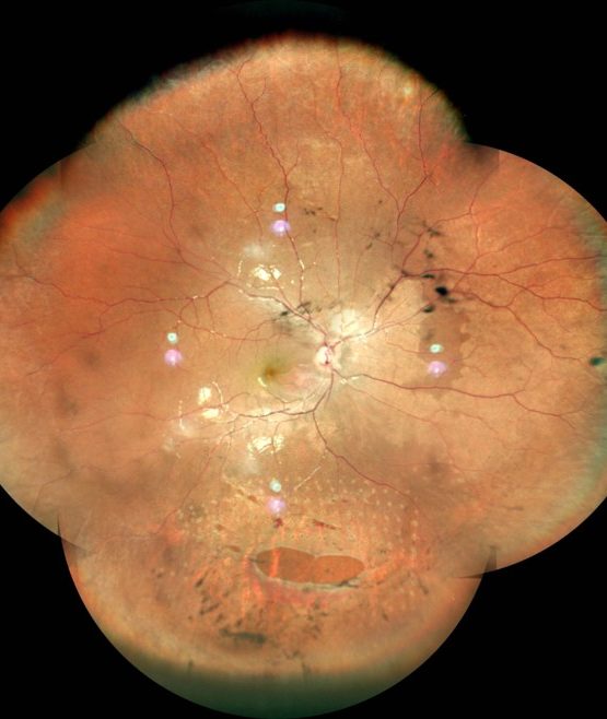
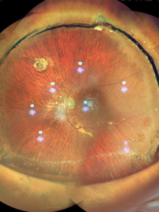
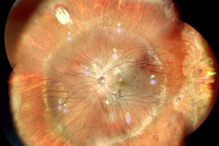
Laser therapy treats lasered lattice and proliferations related to retinal detachments. It creates small burns around tears to seal them and prevent further detachment.
Macular Disorders Captured in Images
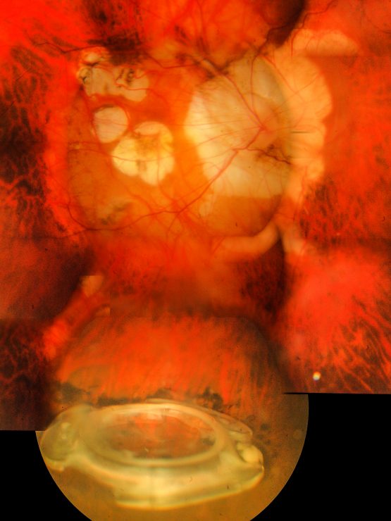
Macular Pucker in Oil-Filled Eye
Causes and Symptoms
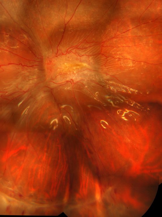
Macular pucker occurs when scar tissue forms on the macula, leading to distorted central vision. In oil-filled eyes, this condition can worsen due to previous surgeries or trauma.
Imaging Insights
Optical Coherence Tomography (OCT) provides high-resolution insights into macular pucker, allowing for precise diagnosis and monitoring.
Traumatic Macula Injuries
Recognizing Trauma Effects
Traumatic injuries to the macula result from blunt force or penetrating trauma. These injuries are recognized through advanced imaging techniques like OCT and fundus photography, revealing structural damage.
Treatment Approaches
Treatment options vary based on severity but may include vitrectomy or laser therapy to repair damaged tissues and restore as much vision as possible.
Macular Hole Diagnosis Using Advanced Imaging Techniques
Diagnosis Process
Diagnosing macular holes involves using OCT and other advanced imaging methods to capture detailed images of the macula’s structure. These images help identify holes early for timely treatment.
Impact on Vision
Untreated macular holes can lead to significant vision loss, making early detection crucial for maintaining visual quality through surgical repair if necessary.
Choroidal and Vascular Conditions
Choroidal Melanoma: Detecting Cancerous Growths Early
Early Detection Importance
Early detection is vital for managing choroidal melanoma effectively. Catching this cancerous growth early improves treatment outcomes significantly.
Imaging Techniques Used
Specific imaging techniques such as fluorescein angiography detect choroidal melanoma by highlighting abnormal blood vessels within the tumor.
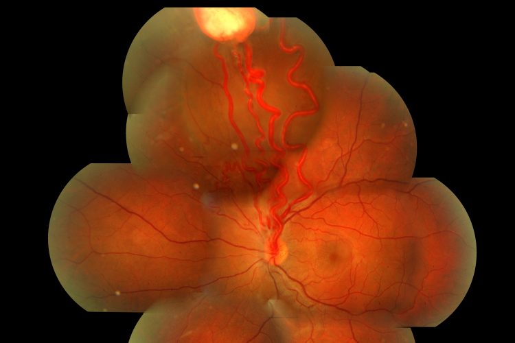
Capillary Haemangioma: Identifying This Vascular Anomaly
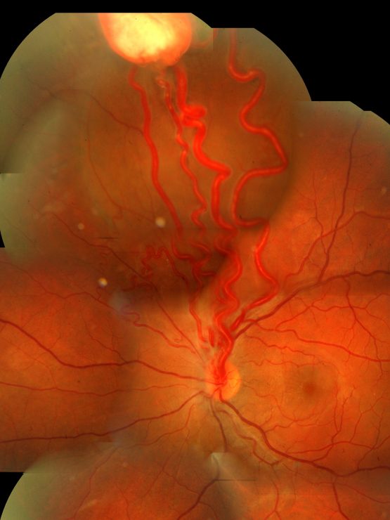
Capillary haemangiomas are benign vascular tumors identified through detailed ocular images using OCT or fundus photography that show characteristic patterns indicative of this anomaly.
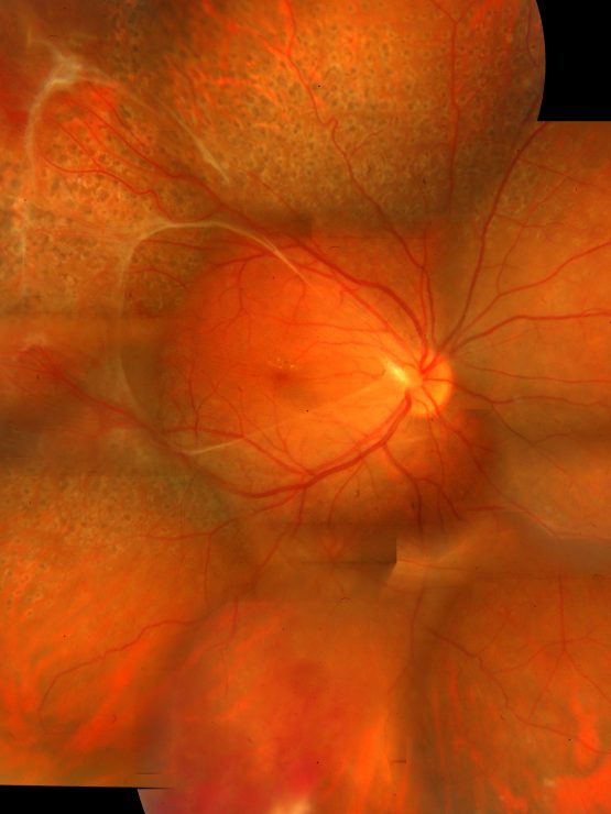
Post-Traumatic Choroidal Striae: Understanding Post-Injury Changes
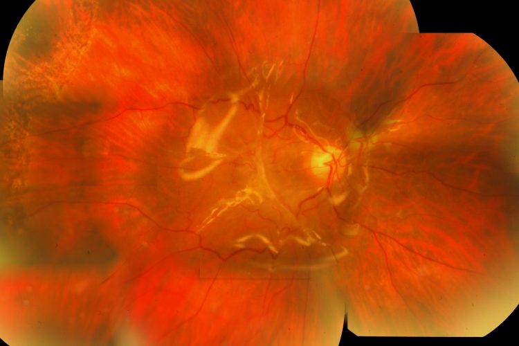
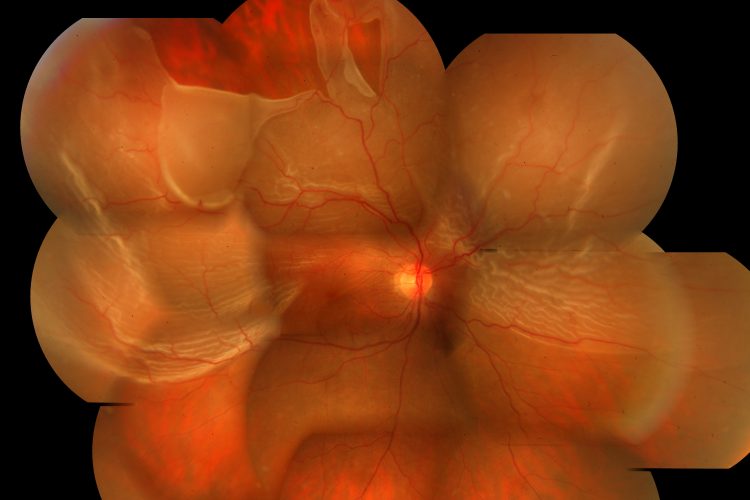
Post-traumatic choroidal striae appear as linear streaks following eye injury. Advanced imaging techniques like OCT or fundus photography provide detailed insights into these changes, aiding diagnosis and management strategies.
Cases of Silicone Oil and Gas Bubble Interference
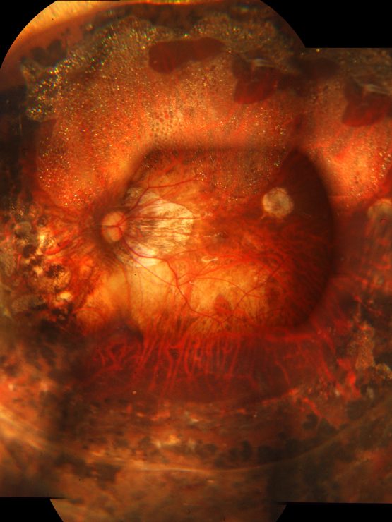
Silicon Oil Filled Eye: Complications And Management
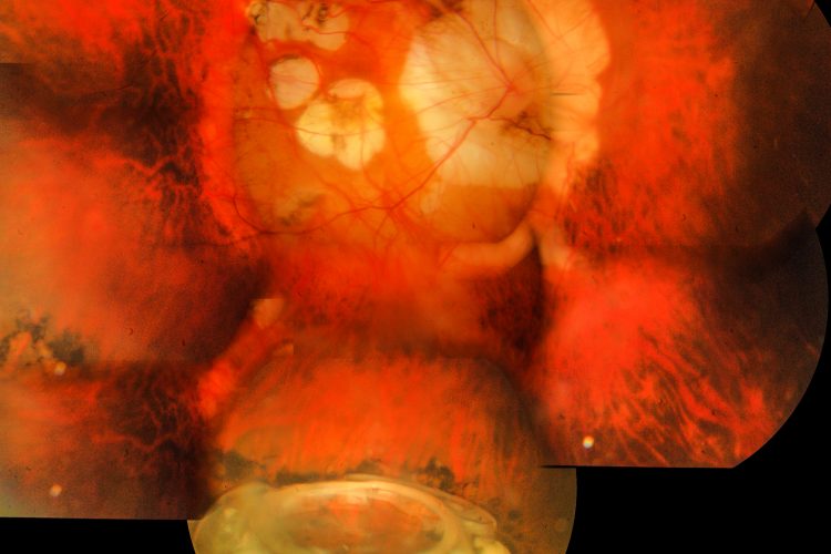
Silicone oil is often used in vitreoretinal surgeries but can lead to complications like emulsification or increased intraocular pressure. Management strategies at Retina Foundation include regular monitoring through imaging techniques and potential surgical interventions if complications arise.
Emulsified Silicon Oil In Subsilicon Space: Challenges In Imaging
Detecting emulsified silicone oil presents challenges due to its scattering properties in ocular media. Modern technological advancements in imaging help overcome these challenges by providing clearer views despite interference from emulsified oil particles.
Inflammatory And Infectious Retinal Conditions
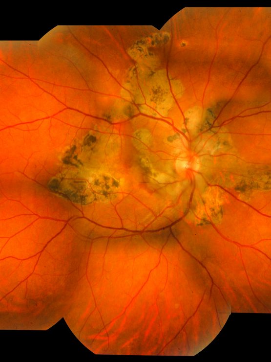
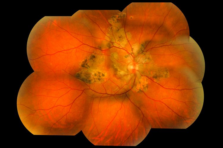
Toxoplasma Scar: Recognizing Infection Related Scarring
Toxoplasma scars result from parasitic infections causing localized scarring on the retina. Recognition involves advanced ocular photographs that highlight characteristic scar patterns aiding accurate diagnosis.
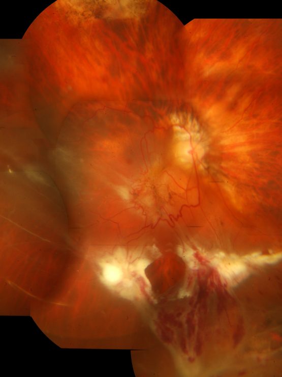
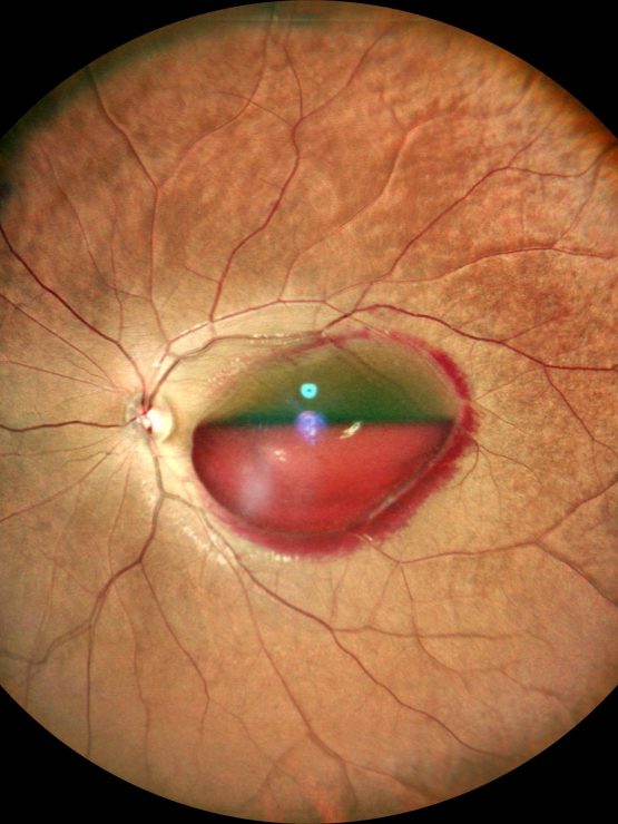
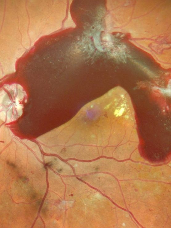
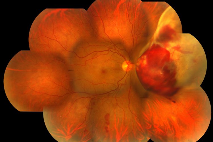
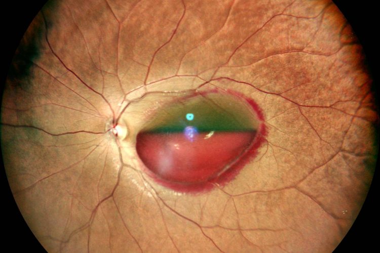
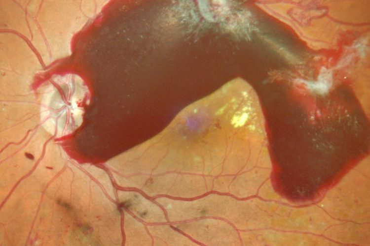
Subhyaloid Hemorrhage: Diagnosing Blood Presence Over The Retina
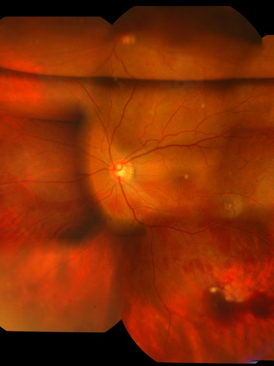
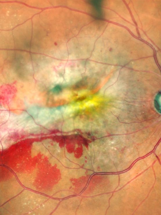
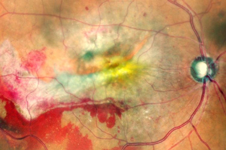
Diagnosing subhyaloid hemorrhage involves capturing digital photographic evidence showing blood accumulation overlying the macula region affecting central vision clarity.
Diagnostic Tools and Techniques in Retinal Imaging
Overview of Retinal Imaging Techniques
Retinal imaging has changed how we look at eye health, giving us detailed views of the retina. Here are some key techniques:
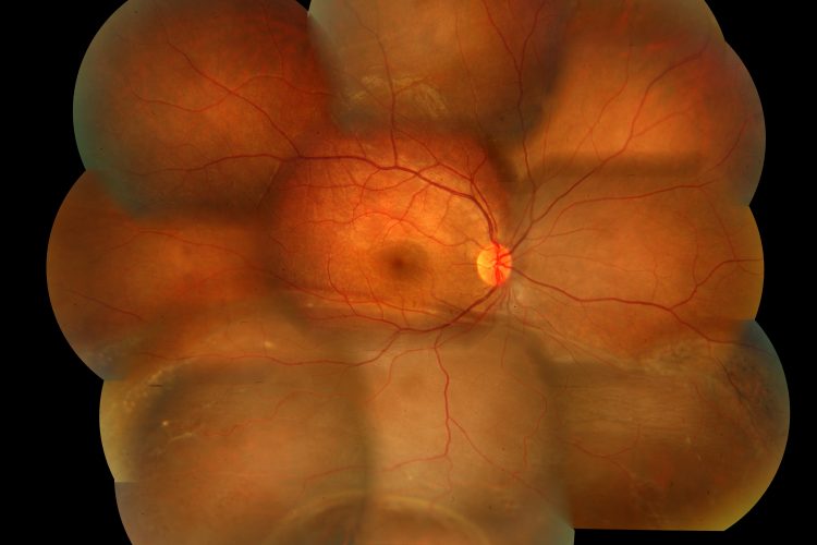
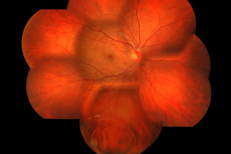
- Fundus Photography: This captures high-resolution images of the inside surface of the eye, including the retina, optic disc, macula, and posterior pole.
- OCT Imaging (Optical Coherence Tomography): Uses light waves to take cross-section pictures of the retina. It helps detect conditions like macular degeneration.
- Fluorescein Angiography: A fluorescent dye is injected into the bloodstream to highlight blood vessels in the retina and spot abnormalities.
- Eye Scans: This term covers various methods used to capture detailed images of different parts of the eye.
- Retinal Exam: A comprehensive evaluation using multiple imaging techniques to assess retinal health.
These techniques provide crucial information through eye retina images that help diagnose various retinal conditions.
How These Techniques Aid Accurate Diagnosis
Advanced retinal imaging techniques are essential for accurate diagnosis. Here’s how they help:
- Central Retinal Artery Occlusion: High-resolution OCT can detect blockages in retinal arteries early on.
- Sub-Retinal Neovascular Membrane: Fluorescein angiography helps identify abnormal blood vessel growth under the retina.
- Macular Pucker: Fundus photography and OCT imaging reveal wrinkles or bulges on the macula’s surface.
- Diabetic Retinopathy Images: Eye scans and fluorescein angiography detect changes due to diabetes, such as hemorrhages or microaneurysms.
- AMD Images (Age-related Macular Degeneration): OCT provides detailed images showing drusen deposits or pigmentary changes associated with AMD.
Using these tools, healthcare professionals can diagnose conditions accurately and start timely treatment plans.
Advantages of Using Advanced Equipment at Retina Foundation
At Retina Foundation, we use advanced technology for better diagnostic outcomes. The benefits include:
- Precise Measurements: State-of-the-art equipment ensures accurate measurements critical for diagnosing and monitoring retinal diseases.
- Clear Imaging: High-definition imaging technologies provide clear visuals that enhance diagnostic accuracy.
- Patient Comfort: Modern equipment is designed to be patient-friendly, ensuring comfort during exams.
- Efficient Diagnosis: Advanced technology allows for quicker diagnosis, enabling prompt treatment initiation.
Our commitment to advanced technology underscores our dedication to providing excellent care.
Ensuring Comprehensive Eye Exams
Comprehensive eye exams are vital for maintaining good eye health. At Retina Foundation, we ensure thorough evaluations by:
- Combining Techniques for an All-Encompassing View
- We integrate multiple imaging methods like fundus photography, OCT imaging, and fluorescein angiography to get a complete picture of retinal health.
- Periodical Monitoring and Follow-Ups
- Regular check-ups using these advanced techniques help track changes over time and adjust treatments accordingly.
This holistic approach ensures no aspect of your eye health is overlooked.
Professional Expertise in Retinal Imaging
The expertise at Retina Foundation sets us apart. Our team comprises highly skilled professionals who excel in interpreting complex retinal images:
- The experienced team at Retina Foundation includes ophthalmologists and technicians specialized in retinal imaging.
- Skilled professionals analyse each image carefully to provide accurate diagnoses and effective treatment plans.
Their proficiency ensures you receive expert care tailored to your specific needs.
Treatment Options and Preventive Care Based on Retinal Imaging
Medical Treatments for Retinal Conditions
Retinal conditions need precise treatments to save vision. Laser therapy is a common choice for lasered lattice and proliferations. This method uses focused laser beams to seal retinal tears or reduce abnormal blood vessel growth. It’s minimally invasive and usually done in an outpatient setting.
Vitrectomy is a more complex surgery, used when there are issues like retinal detachment, macular holes, or macular puckers. In this procedure, the vitreous gel is removed and replaced with a saline solution or gas bubble to help reattach the retina. Managing emulsified silicon oil after vitrectomy is crucial to avoid further complications.
Specific conditions like central retinal artery occlusion and subhyaloid hemorrhage need tailored medical interventions. For example, immediate treatment for central retinal artery occlusion might involve ocular massage or medications to lower intraocular pressure.
Lifestyle Adjustments for Better Eye Health
Maintaining good eye health involves several lifestyle changes:
- Diet and Nutrition Tips for Eye Health: Eat foods rich in antioxidants, vitamins A, C, E, and omega-3 fatty acids. Leafy greens, fish like salmon, nuts, and citrus fruits are especially beneficial.
- Reducing Screen Time Impact on Retinal Health: Limit screen time by taking regular breaks using the 20-20-20 rule—every 20 minutes, look at something 20 feet away for at least 20 seconds. Adjust your screen brightness to match ambient lighting.
- Exercises to Support Eye Strength: Do eye exercises such as focusing on distant objects periodically throughout the day. Simple routines like rolling your eyes in different directions can also help maintain eye muscle flexibility.
Preventive Strategies at Retina Foundation
Preventive care is key at Retina Foundation. Regular screenings and check-ups are vital for early detection of retinal conditions. These screenings often include comprehensive eye exams that use advanced imaging technologies to spot abnormalities before symptoms appear.
Personalized care plans are developed based on detailed imaging results from these screenings. Each plan addresses specific needs identified through high-resolution images of the retina. This proactive approach ensures potential issues are managed before they become serious problems.
Utilizing Retinal Images for Effective Treatment Plans
Retinal images play a crucial role in creating effective treatment plans. The process starts with a detailed diagnosis using high-quality images that show the condition’s specifics.
From diagnosis to treatment roadmap:
- Initial Diagnosis: High-resolution imaging identifies problem areas.
- Treatment Planning: Based on imaging results, a step-by-step treatment plan is created.
- Implementation: Treatments such as laser therapy or vitrectomy are carried out according to the plan.
- Follow-Up Imaging: Post-treatment images ensure the interventions were effective.
Case studies showcasing successful treatments initiated from detailed imaging highlight how precise diagnostics lead to better patient outcomes.
Future Trends in Retinal Imaging and Treatments
The future of retinal care looks bright with new technologies in retinal photography set to change eye care:
- Emerging Technologies: Innovations like adaptive optics scanning laser ophthalmoscopy (AOSLO) offer unmatched detail in retinal imaging.
- Future Advancements: Artificial intelligence (AI) integration into diagnostic tools predicts disease progression more accurately than ever before.
These advancements not only improve diagnostic accuracy but also open new ways for personalized treatment options based specifically on individual patient data obtained through advanced imaging techniques.
Frequently Asked Questions and Expert Advice
Common Concerns Regarding Retinal Imaging
Retinal photography is a safe and non-invasive procedure that captures detailed images of the eye retina. Techniques like fundus photography, OCT imaging, and fluorescein angiography help detect and monitor various eye conditions without direct contact with the eye, ensuring patient comfort.
How often you need a retinal exam depends on factors like age, family history of eye diseases, and health conditions such as diabetes. Generally, it’s recommended to have a retinal exam every one to two years. During a retinal imaging session, patients can expect a quick and painless process where their eyes are dilated for better visualization of the retina.
Expert Tips for Maintaining Optimal Eye Health
- UV Protection: Wearing sunglasses with UV protection shields your eyes from harmful ultraviolet rays.
- Proper Eyewear: Using appropriate eyewear for activities like reading or working on computers reduces strain on your eyes.
- Diet and Nutrition: Eating foods rich in vitamins A, C, E, and omega-3 fatty acids supports overall eye health. Leafy greens, fish, nuts, and citrus fruits are excellent choices.
- Reducing Screen Time: Limiting screen time and taking regular breaks using the 20-20-20 rule (every 20 minutes, look at something 20 feet away for at least 20 seconds) helps reduce digital eye strain.
- Exercises for Eye Strength: Simple exercises like focusing on distant objects or performing eye rotations can enhance eye muscle strength.
Myths and Facts About Retinal Imaging
There are several misconceptions about retinal imaging:
- Myth: Retinal imaging is painful.
Fact: Retinal photography is completely painless and non-invasive. - Myth: Only older adults need retinal exams.
Fact: Regular retinal exams are important for individuals of all ages to detect early signs of conditions like glaucoma or diabetic retinopathy. - Myth: Retinal imaging exposes you to harmful radiation.
Fact: Techniques like OCT imaging use light waves instead of radiation to capture detailed images of the retina.
Understanding these facts helps appreciate the true benefits of retinal photography in maintaining good ocular health.
How Retina Foundation Stands Out
Retina Foundation stands out through its commitment to patient care and use of advanced technology in retinal imaging. Employing techniques such as fundus photography and OCT imaging ensures precise diagnoses. The experienced team provides personalized attention tailored to each patient’s needs.
The foundation continually invests in state-of-the-art equipment and ongoing staff training to stay at the forefront of ophthalmic care. This dedication ensures high-quality service delivery that sets them apart from other providers.
Real-life Testimonials
Patients who have undergone detailed retinal imaging at Retina Foundation often share transformative stories:
- John D.: “Early detection through OCT imaging saved my vision by identifying macular degeneration before it progressed.”
- Priya S.: “The comprehensive care I received helped manage my diabetic retinopathy effectively.”
- Ravi K.: “Regular fundus photography sessions allowed timely intervention for my glaucoma.”
These testimonials highlight how advanced retinal imaging has positively impacted lives by enabling early diagnosis and effective treatment plans tailored by Retina Foundation’s expert team.
Summary of Key Points
Advanced retinal photography is crucial for maintaining eye health. Techniques like OCT imaging and fundus photography can detect serious conditions such as retinal detachment, macular hole, and macular pucker. These technologies offer detailed eye retina images that help in diagnosing retinal pathologies accurately. Early detection of issues like retinal artery occlusion, choroidal melanoma, and retinitis pigmentosa through these ocular images ensures timely treatment planning and better outcomes.
Encouraging Regular Eye Check-ups
Scheduling regular eye check-ups at Retina Foundation is vital for early detection of eye conditions. Routine screenings using advanced imaging techniques like fundus photography and OCT imaging can identify problems such as diabetic retinopathy and age-related macular degeneration early on. Regular monitoring with these tools helps in managing these conditions effectively, preserving vision and overall eye health.
Final Thoughts on Retinal Imaging Advancements
The future of retinal imaging is promising with emerging technologies like 3D eye imaging and digital retinal photography. These advancements are set to revolutionise how we monitor ocular health, offering more precise diagnostics and better treatment options. Optical coherence tomography (OCT) and fluorescein angiography are already making significant strides in this field.


